
The National Institute of Biomedical Imaging and Bioengineering (NIBIB) supports a large network of National Centers for Biomedical Imaging and Bioengineering (NCBIB) through the P41 grant mechanism. These Centers create critical and unique technology and methods at the forefront of their respective fields and apply them to a broad range of basic, translational and clinical research. This occurs through a synergistic interaction of technical and biomedical expertise, both within the Centers and in intensive collaborations with other leading laboratories. Scientists at these Centers ensure that NIH-funded biomedical research projects may gain access to the newest and most advanced technologies, techniques, and methodologies. The National Institute of General Medical Sciences (NIGMS) has a similar program. Details about that program can be found at Biomedical Technology Development and Dissemination Research Centers (BTDD). Applicants who are interested in submitting an application to the NIGMS program need to use NIGMS application procedures rather than those in this announcement.
NIBIB Supported Centers
View NIBIB-supported National Centers for Biomedical Imaging and Bioengineering
Application Information
- Guidelines for NIBIB P41 National Centers for Biomedical Imaging and Bioengineering (NCBIB) Adobe PDF (119KB)
- Requirements for a “New” NIBIB P41 National Center Following Funding Period Limitations Adobe PDF (114KB)
- Current Program Announcement (PAR-20-169)
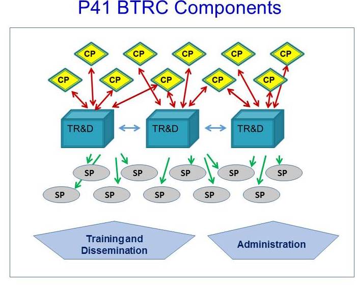
- NIBIB P41 NCBIB Components (JPG Image (50K)
- Using ASSIST to prepare and submit multi-project application to NIH Adobe PDF (23MB)
- "Play" with ASSIST for applicants Adobe PDF (125K)
- Download P41 White Paper Template Word DOCX (53KB) | Instructions for completing white paper
- Download P41 White Paper Budget Table Template (Excel Worksheet 35KB)
NCBIB Progress Report Information
Webinars
The National Technology Centers (NCBIB) Webinar Series showcases the innovative work of NIBIB-funded centers. Register now and join us for the next webinar.