Advances in Heart Health Research
Download PDF
NIBIB Researching the Heart.pdf
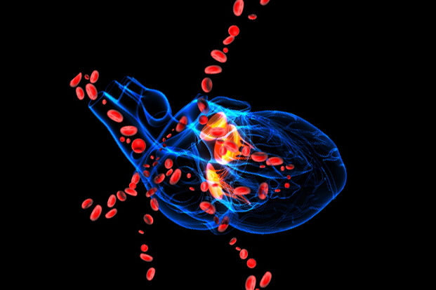
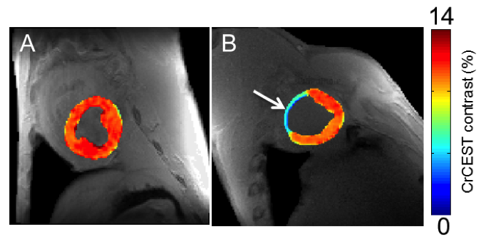
Revealing Heart Damage Earlier
Researchers funded by the National Institutes of Health have developed a high-resolution MRI technique to visualize disruptions in energy metabolism in the heart, often a sign of disease. The technique could help doctors identify damaged heart tissue earlier in patients with coronary artery disease or other heart disorders.
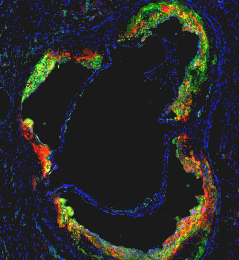
Reducing Recurrent Heart Attacks and Stroke
Up to 30 percent of heart attack patients suffer a new heart attack because cardiologists are unable to control inflammation inside heart arteries — the process that leads to clots rupturing and causing myocardial infarction or stroke.
An international research team, led by Mount Sinai investigators, designed and tested a high-density lipoprotein (HDL) nanoparticle loaded with a statin drug. In mouse studies, they show this HDL nanotherapy is capable of directly targeting and lowering dangerous inflammation in blood vessels. Read more...
Source: Mount Sinai Hospital
Resorbable Cardiovascular Stent
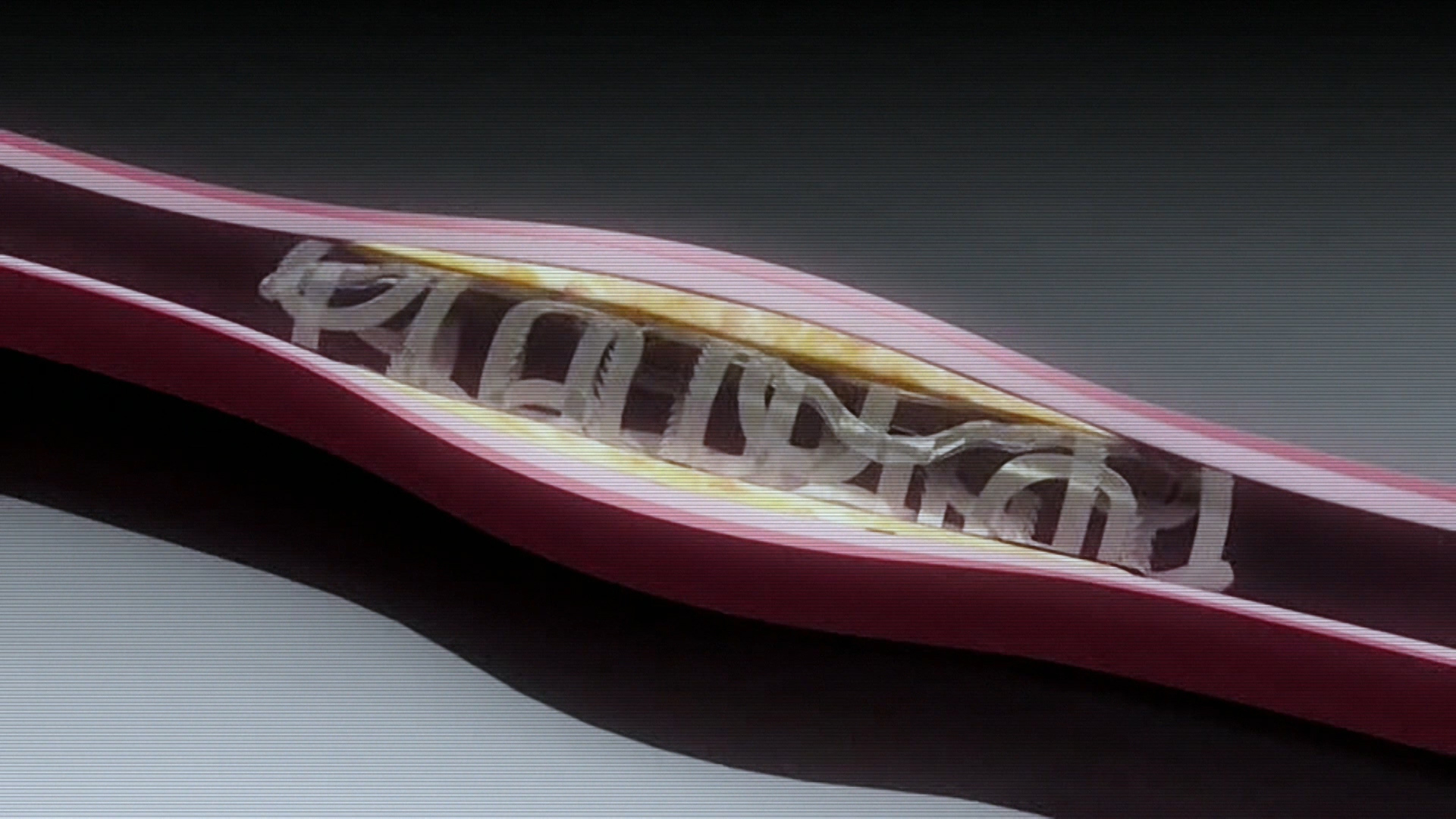
NIH funded research at Rutgers University has resulted in the development of new materials for biomedical applications. One example is a new resorbable cardiovascular stent that can replace the metal stents currently in use, which remain in the body and often cause long-term complications. This stent also delivers medication and is radio-opaque, allowing imaging and controlled monitoring of the re-absorption process. Read more...
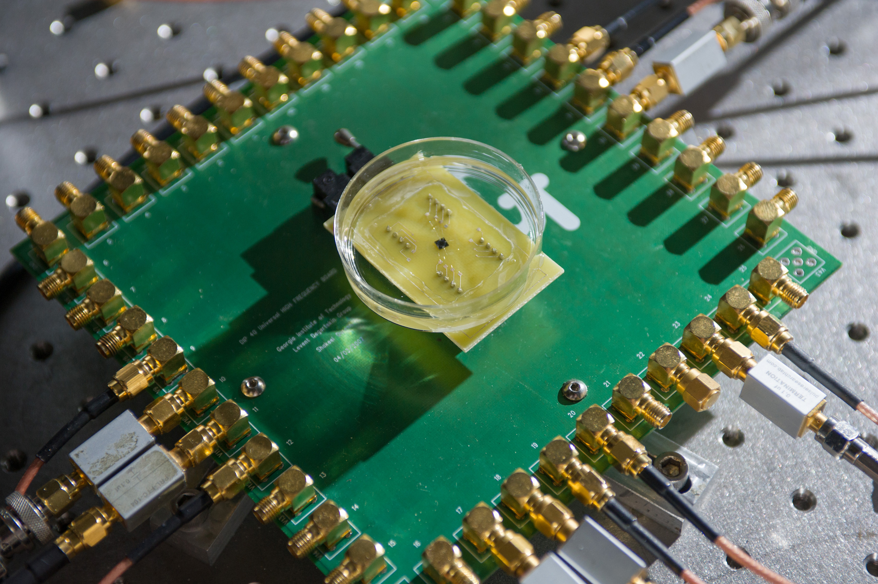
Chip Images the Heart from Inside
Researchers have developed the technology for a catheter-based device that would provide forward-looking, real-time, three-dimensional imaging from inside the heart, coronary arteries and peripheral blood vessels. With its volumetric imaging, the new device could better guide surgeons working in the heart, and potentially allow more of patients’ clogged arteries to be cleared without major surgery.
Source: Georgia Institute of Technology