
- What is a computed tomography (CT) scan?
- How does CT work?
- When would I get a CT scan?
- What is a CT contrast agent?
- Are there risks?
- What are examples of NIBIB-funded projects using CT?
What is a computed tomography (CT) scan?
The term “computed tomography,” or CT, refers to a computerized x-ray imaging procedure in which a narrow beam of x-rays is aimed at a patient and quickly rotated around the body, producing signals that are processed by the machine’s computer to generate cross-sectional images, or “slices.” These slices are called tomographic images and can give a clinician more detailed information than conventional x-rays. Once a number of successive slices are collected by the machine’s computer, they can be digitally “stacked” together to form a three-dimensional (3D) image of the patient that allows for easier identification of basic structures as well as possible tumors or abnormalities.
How does CT work?
Unlike a conventional x-ray—which uses a fixed x-ray tube—a CT scanner uses a motorized x-ray source that rotates around the circular opening of a donut-shaped structure called a gantry. During a CT scan, the patient lies on a bed that slowly moves through the gantry while the x-ray tube rotates around the patient, shooting narrow beams of x-rays through the body. Instead of film, CT scanners use special digital x-ray detectors, which are located directly opposite the x-ray source. As the x-rays leave the patient, they are picked up by the detectors and transmitted to a computer.

Each time the x-ray source completes one full rotation, the CT computer uses sophisticated mathematical techniques to construct a two-dimensional image slice of the patient. The thickness of the tissue represented in each image slice can vary depending on the CT machine used, but usually ranges from 1-10 millimeters. When a full slice is completed, the image is stored and the motorized bed is moved forward incrementally into the gantry. The x-ray scanning process is then repeated to produce another image slice. This process continues until the desired number of slices is collected.
Image slices can either be displayed individually or stacked together by the computer to generate a 3D image of the patient that shows the skeleton, organs, and tissues as well as any abnormalities the physician is trying to identify. This method has many advantages including the ability to rotate the 3D image in space or to view slices in succession, making it easier to find the exact place where a problem may be located.
When would I get a CT scan?
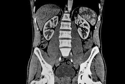
CT scans can be used to identify disease or injury within various regions of the body. For example, CT has become a useful screening tool for detecting possible tumors or lesions within the abdomen. A CT scan of the heart may be ordered when various types of heart disease or abnormalities are suspected. CT can also be used to image the head in order to locate injuries, tumors, clots leading to stroke, hemorrhage, and other conditions. It can image the lungs in order to reveal the presence of tumors, pulmonary embolisms (blood clots), excess fluid, and other conditions such as emphysema or pneumonia. A CT scan is particularly useful when imaging complex bone fractures, severely eroded joints, or bone tumors since it usually produces more detail than would be possible with a conventional x-ray.
What is a CT contrast agent?
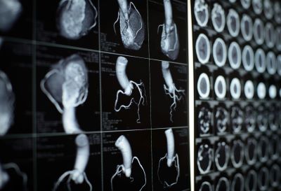
As with all x-rays, dense structures within the body—such as bone—are easily imaged, whereas soft tissues vary in their ability to stop x-rays and therefore may be faint or difficult to see. For this reason, contrast agents have been developed that are highly visible in an x-ray or CT scan and are safe to use in patients. Contrast agents contain substances that can stop x-rays and are therefore more visible on an x-ray image. For example, to examine the circulatory system, an intravenous (IV) contrast agent based on iodine is injected into the bloodstream to help illuminate blood vessels. This type of test is used to look for possible obstructions in blood vessels, including those in the heart. Oral contrast agents, such as barium-based compounds, are used for imaging the digestive system, including the esophagus, stomach, and gastrointestinal (GI) tract.
Are there risks?
CT scans can diagnose possibly life-threatening conditions such as hemorrhage, blood clots, or cancer. An early diagnosis of these conditions could potentially be lifesaving. However, CT scans use x-rays, and all x-rays produce ionizing radiation. Ionizing radiation has the potential to cause biological effects in living tissue. This is a risk that increases with the number of exposures added up over the life of an individual. However, the risk of developing cancer from x-ray radiation exposure is generally small.
A CT scan in a pregnant woman poses no known risks to the baby if the area of the body being imaged isn’t the abdomen or pelvis. In general, if imaging of the abdomen and pelvis is needed, doctors prefer to use exams that do not use radiation, such as magnetic resonance imaging (MRI) or ultrasound. However, if neither of those can provide the answers needed, or there is an emergency or other time constraint, CT may be an acceptable alternative imaging option.
In some patients, contrast agents may cause allergic reactions, or in rare cases, temporary kidney failure. IV contrast agents should not be administered to patients with abnormal kidney function since they may induce a further reduction of kidney function, which may sometimes become permanent.
Because children are more sensitive to ionizing radiation and have a longer life expectancy, they have a higher relative risk for developing cancer from such radiation compared with adults. Parents may want to ask the technologist or doctor if their machine settings have been adjusted for children.
What are examples of NIBIB-funded projects using CT?
New and improved CT receives FDA clearance
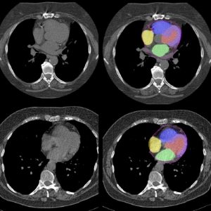
Researchers at Mayo Clinic have developed the first photon-counting detector (PCD)-CT system, which, unlike traditional CT, measures individual x-rays as they leave a patient, allowing for more detailed medical images. This system offers improved resolution and signal, reduces the overall radiation dose, and lowers the amount of contrast agent, which can pose risks to certain patients, needed for CT imaging. The team demonstrated their method’s capabilities by comparing conventional CT to PCD-CT scans in several clinical studies. The first PCD-CT imaging device was cleared by the U.S. Food and Drug Administration (FDA) in 2021.
Using AI to repurpose routine CT scans
Heart disease is largely preventable, yet it is the leading cause of death in the U.S., and many people don’t understand their risk until it is too late. To increase the accessibility of early cardiac screening, researchers at Cedars-Sinai Medical Center are developing an artificial intelligence (AI) tool that repurposes standard chest CT scans, often performed for lung-related conditions, to predict mortality. In a recent study, the team evaluated several types of CT scans and successfully identified cardiac factors predictive of death in thousands of patients. This opportunistic screening method could potentially flag at-risk patients for follow-up scans or life-saving interventions.
Updated July 2025