3D Retinal Tissue Bioreactor for Retinal Therapy Development

Share:

EmailFacebookLinkedInXWhatsAppShare

Collaborators

Neurobiology Neurodegeneration & Repair Laboratory, NEI
Biomedical Engineering & Physical Science Shared Resource Program, NIBIB

Project Brief

Retinal degenerative diseases currently have limited treatment options partly due to a lack of effective in vitro model systems to investigate mechanisms of disease pathogenesis and evaluate new therapies. To face this challenge, research has been conducted to culture three-dimensional (3D) retinal organoids from mammalian pluripotent stem cells (PSC). In static culture, PSC-derived neural retina (NR) mimics in vivo mouse retina with proper polarity and limited stratification of major cell types.  However, these NRs are still limited by the immaturity of their photoreceptors, which do not develop the outer segment structures necessary for capturing light photons. In in vivo retina, outer segments are embedded in the retinal pigment epithelium (RPE) for structural support and nutrient transport; thus, co-culturing NR with RPE tissue may further differentiate photoreceptors. The proposed retinal organoids may generate photoreceptor cells suitable for cell replacement therapies that could potentially restore vision. Furthermore, taking advantage of the indefinite proliferation and capacity to differentiate into various tissue types, this co-culture technology for PSC-derived organoids could enhance the methodology investigators use to study disease pathogenesis in other organ systems.

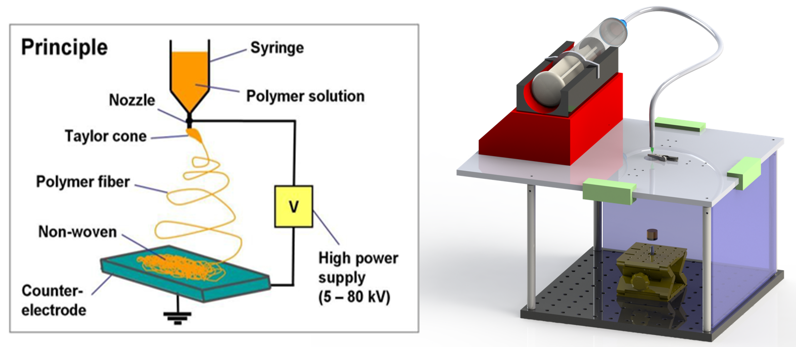
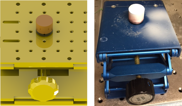
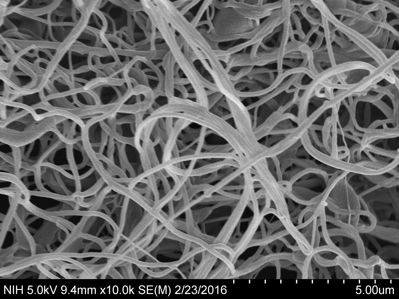
Bruch’s membrane is a 2-4 µm thick extracellular matrix located between the RPE and choroid layers.  The membrane provides structural support and facilitates nutrient transfer to the RPE. The membrane has major clinical significance because of its role in retinal degenerative diseases, such as age-related macular degeneration (AMD) and retinitis pigmentosa (RP). In collaboration with NEI and NIBIB, SPIS engineers are developing an electrospinning system to generate a nanofibrous scaffold material from poly (ε-caprolactone) (PCL).  The PCL nanofibrous scaffold will be used to culture a PSC-derived RPE monolayer.

Computer generated diagram of the device and how the 3D printer works with a nozzle and orange string on the image on the left. The image on the left is a small red box on top with a wire going down to a larger clear box.

Diagram of electrospinning principle and 3D drawing of the electrospinning system setup.  A syringe pump delivers a polymer solution (at a constant flow rate) to a hypodermic needle attached to a high-voltage (5-80 kV) DC power supply. The high voltage applied to the liquid droplet causes the polymer to elongate into a filament due to electrostatic repulsion towards the ground potential collector, whereupon the fiber is deposited in random orientations to form a fibrous mat.  The elongation results in the formation of fibers with diameters in the nanometer range.


A CAD diagram (yellow) and photo (blue) of the nanofiber collector device.

3D CAD model and photo of the grounded nanofiber collector device. The nanofiber was collected for ~30 minutes.  The fiber accumulated uniformly over the entire top surface of the collector to form the fibrous scaffold material (white material on top of the copper cylindrical piece) from poly (ε-caprolactone) (PCL).


A black and white electron microscopy image of long strands

Scanning electron microscopy image of electrospun nano-fibrous scaffold material from poly (ε-caprolactone) (PCL). The fiber was generated from a 12mg/mL PCL in hexafluoro-2-propanol (HFIP) polymer concentration with the following electrospinning system settings:  5kV high voltage, 1mL/hr polymer flowrate, and 12cm distance from needle tip to collector top.