
- What are medical x-rays?
- How do medical x-rays work?
- When are medical x-rays used?
- Are there risks?
- What are NIBIB-funded researchers developing in the field of x-ray technology?
What are medical x-rays?
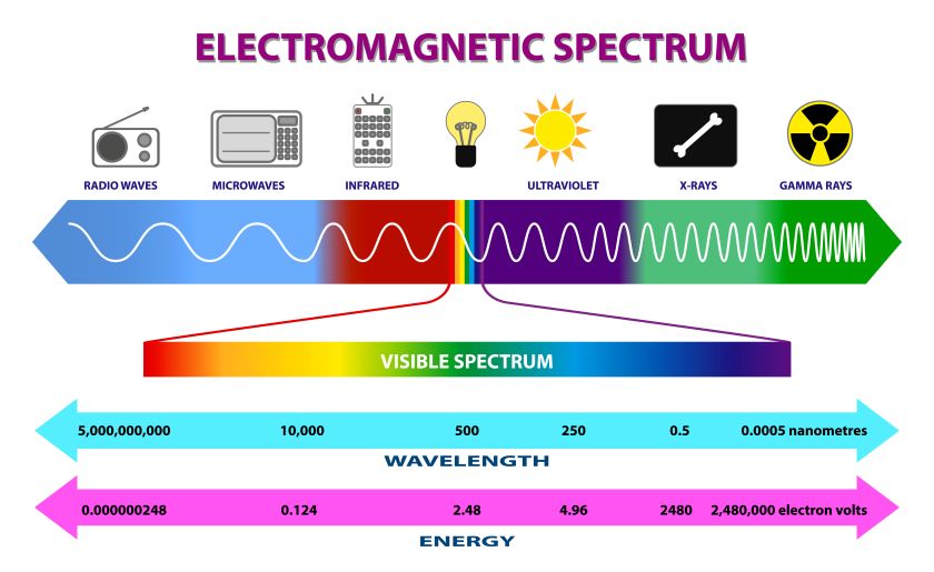
X-rays are a form of electromagnetic radiation, similar to visible light. Unlike light, however, x-rays have higher energy and can pass through most objects, including the body. Medical x-rays are used to generate images of tissues and structures inside the body. If x-rays traveling through the body also pass through an x-ray detector on the other side of the patient, an image will be formed that represents the “shadows” formed by the objects inside of the body.
One type of x-ray detector is photographic film, but there are many other types of detectors that are used to produce digital images. The x-ray images that result from this process are called radiographs.
How do medical x-rays work?
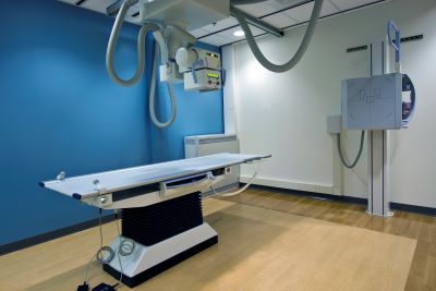
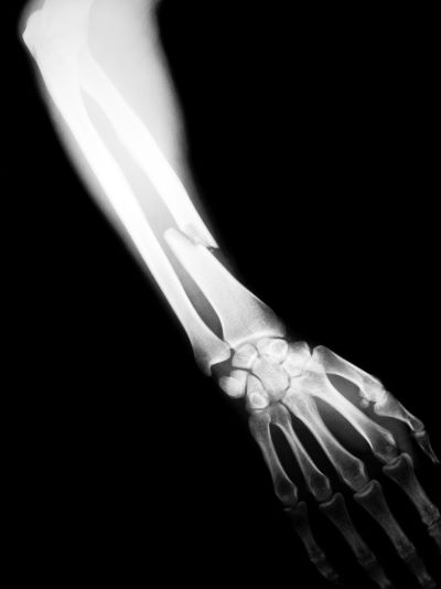
To create a radiograph, a patient is positioned so that the part of the body being imaged is located between an x-ray source and an x-ray detector. When the machine is turned on, x-rays travel through the body and are absorbed in different amounts by different tissues, depending on the radiological density of the tissues they pass through. Radiological density is determined by both the density and the atomic number (the number of protons in an atom’s nucleus) of the material being imaged. For example, our bones contain calcium, which has a higher atomic number than most other tissues. Because of this property, bones readily absorb x-rays and therefore produce high contrast on the x-ray detector. As a result, bony structures appear whiter than other tissues against the black background of a radiograph. Conversely, x-rays travel more easily through less radiologically dense tissues, such as fat, muscle, and air-filled cavities such as the lungs. These structures are displayed in shades of gray on a radiograph.
When are medical x-rays used?
Listed below are examples of examinations and procedures that use x-ray technology to either diagnose or treat disease:
Diagnostic
X-ray radiography: Detects bone fractures, certain tumors and other abnormal masses, pneumonia, some types of injuries, calcifications, foreign objects, or dental problems.
Mammography: A radiograph of the breast that is used for cancer detection and diagnosis. Tumors tend to appear as regular or irregular-shaped masses that are somewhat brighter than the background on the radiograph (i.e., whiter on a black background or blacker on a white background). Mammograms can also detect tiny bits of calcium, called microcalcifications, which show up as very bright specks on a mammogram. While usually benign, specific patterns of microcalcifications could indicate the presence of cancer. Learn more about mammography here.
Computed tomography (CT): Combines traditional x-ray technology with computer processing to generate a series of cross-sectional images of the body that can later be combined to form a three-dimensional x-ray image. CT images are more detailed than plain radiographs and give doctors the ability to view structures within the body from many different angles. Learn more about CT here.
Fluoroscopy: Uses x-rays and a fluorescent screen to obtain real-time images of movement within the body or to view diagnostic processes, such as following the path of an injected or swallowed contrast agent. For example, fluoroscopy is used to view the movement of the beating heart, and, with the aid of radiographic contrast agents, to view blood flow to the heart muscle as well as through blood vessels and organs. This technology is also used with a radiographic contrast agent to guide an internally threaded catheter during cardiac angioplasty, which is a minimally invasive procedure for opening clogged arteries that supply blood to the heart.
Therapeutic
Radiation therapy in cancer treatment: X-rays and other types of high-energy radiation can be used to destroy cancerous tumors and cells by damaging their DNA. The radiation dose used for treating cancer is much higher than the radiation dose used for diagnostic imaging. Therapeutic radiation can come from a machine outside of the body or from a radioactive material that is placed in the body, inside or near tumor cells, or injected into the blood stream. Learn more about radiation treatment for cancer therapy here.
Are there risks?
When used appropriately, the diagnostic benefits of x-ray scans significantly outweigh the risks. X-ray scans can diagnose possibly life-threatening conditions such as blocked blood vessels, bone cancer, and infections. However, x-rays produce ionizing radiation—a form of radiation that has the potential to harm living tissue. This is a risk that increases with the number of exposures added up over the life of an individual. However, the risk of developing cancer from radiation exposure is generally small.
An x-ray in a pregnant woman poses no known risks to the baby if the area of the body being imaged isn’t the abdomen or pelvis. In general, if imaging of the abdomen and pelvis is needed, doctors prefer to use exams that do not use radiation, such as magnetic resonance imaging (MRI) or ultrasound. However, if neither of those can provide the answers needed, or there is an emergency or other time constraint, an x-ray may be an acceptable alternative imaging option.
Because children are more sensitive to ionizing radiation and have a longer life expectancy, they have a higher relative risk for developing cancer from such radiation compared with adults. Parents may want to ask the technologist or doctor if their machine settings have been adjusted for children.
Learn more about specific risks involved with CT and mammography.
What are NIBIB-funded researchers developing in the field of x-ray technology?
Current research of x-ray technology focuses on ways to reduce radiation dose, improve image resolution, and enhance contrast materials and methods. The following are examples of research projects funded by NIBIB that are developing new applications of x-ray technology:
Single-frame x-ray tomosynthesis (SFXT): Conventional x-ray radiography generates a single two-dimensional image, which is created by imaging a single plane at a single time point. X-ray tomosynthesis, on the other hand, uses multiple images, which are then reconstructed to generate more information, such as a three-dimensional image. Unlike CT imaging, where the source/detector physically travels at least 180 degrees around the patient, tomosynthesis uses a limited rotational angle and takes fewer images (requiring less radiation and less expense). Current tomosynthesis approaches, however, generate a static snapshot of the tissue of interest and do not allow for real-time imaging.
NIBIB-funded researchers are working on a new x-ray method, called single-frame x-ray tomosynthesis (or SFXT), that would allow for real-time monitoring of a small area of tissue. By capturing 30 images every second, this technique would have 10 to 100 times the temporal resolution of conventional tomosynthesis, resulting in “sharper” images of tissues in motion (similar to using a faster shutter speed on a camera). The researchers plan to evaluate the use of SFXT in the detection of cardiovascular disease — by looking at calcium deposits in the coronary arteries — and to guide radiation treatment to precise locations in the lungs, which would enable safer ablation of lung tumors.
Imaging to guide lung biopsies: Lung cancer is the leading cause of cancer-related mortality in the United States, and analyzing lesions found in the lungs is a way to screen for the disease and to guide treatment. For a biopsy, one method to obtain lung tissue is through a bronchoscopy, where a thin tube is passed through the nose or mouth and guided into the lungs. However, obtaining tissues of interest remains difficult, as locating and visualizing such lesions is challenging. To overcome these limitations, researchers have developed a new, cost-effective chest x-ray tomosynthesis system that can generate high-resolution, real-time images of the lungs, which would allow for improved visualization during a transbrochial biopsy. In addition to being less expensive and easier to use than standard CT-based approaches, this x-ray technique is stationary and does not require any physical motion of the x-ray source or detector. Further, this method uses low doses of radiation, which would be beneficial for patients who require multiple biopsies. This x-ray system is currently being optimized for pre-clinical large animal evaluation.
Learn more about how X-rays work here.
Updated June 2022