Collaborators
Project Brief
Functional neuroimaging, particularly functional MRI (fMRI) which provides a measure of regional brain blood flow during behavior, is widely used in the investigation of cognitive processes and has been proposed as a means of evaluating deficits due to traumatic brain injury (TBI) and guiding rehabilitation. fMRI, however, requires specialized permanently-sited facilities, as well as specialized personnel, and is not commonly available. There is need for a simpler, easily accessible, low-cost device capable of identifying and potentially imaging hematomas. This hematoma detector device will provide for rapid identification and triage of hematomas with a handheld device useable by a non-specialist. The device will be able to detect mono- and bi-lateral hematomas, and can be designed for portability (i.e. field use).
The proposed optical-based device is moved across the head to detect a hematoma by using the changing optical signal as a differential measure. A single set of fibers, one source and one detector, of fixed physical separation, will certainly provide varying signals corresponding to heterogeneity of the head. However, if two detectors are used—one to probe the depth that includes skin and skull and one to probe the dural region—we can normalize for changes due to superficial heterogeneity. This assumption is valid because we are not, as typical in near-infrared imaging (NIR), looking for hemodynamic changes, but rather detecting the presence of a large inclusion of blood in the dural region. The presence of a localized volume of blood (i.e., hematoma) should provide a larger than normal change in absorption due to the high concentration of blood relative to the surrounding tissue. NIR imaging typically measures changes in blood volume/oxygenation at a blood concentration of around 3% in brain. With our hematoma detector, we aim to measure the difference signal between this normal 3% level and the ~100% concentration in a significantly sized region of the hematoma.
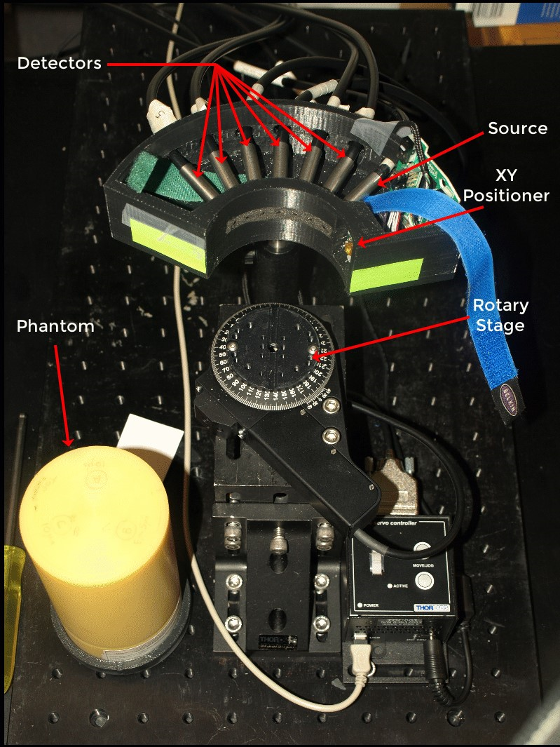
To test the hematoma detector concept using a cylindrical phantom, SPIS staff developed the test fixture that included one source fiber and six detector fibers. The source fiber is connected to four optically coupled laser diodes (800, 850, 900, and 950 nm). The detector fibers are input to six photomultiplier tubes. This optical configuration selected to simultaneously monitor six different depths. With the detector fibers at varying distances from the source fiber, the detected signal amplitudes vary over a large dynamic range. SPIS staff designed and built a custom multichannel amplifier instrument enabling collaborators to independently adjust the gain and offset of each detector channel. SPIS was also responsible for developing the data acquisition system for this prototype system. Using National Instruments hardware and LabVIEW software, we developed a flexible test and validation system enabling phantom studies and downstream algorithm development.