An official website of the United States government
Here’s how you know
Official websites use .gov A
.gov
website belongs to an official government organization in the United States.
Secure .gov websites use HTTPS A
lock
(
) or
https://
means you’ve safely connected to the .gov website. Share sensitive information only on official, secure websites.
High-resolution Gamma Imager for Small Animal Imaging of Radioisotopes for Cancer Treatment
Collaborators
Peter Choyke, M.D.
Molecular Imaging Program, Center for Cancer Research, NCI
Jurgen Seidel, Ph.D.
Molecular Imaging Program, Center for Cancer Research, NCI
Michael Green, Ph.D.
Molecular Imaging Program, Center for Cancer Research, NCI
Wenze Xi, Ph.D.
Radiation Detector and Medical Imaging Group, Thomas Jefferson National Accelerator Facility
Project Brief
SPIS collaborated with NCI’s Molecular Imaging Program (MIP) to develop novel imaging systems used in the development of radionuclide-labeled compounds. Successfully developed radionuclide-labeled compounds offer the ultimate prospect of PET, SPECT and planar imaging in human subjects for medical diagnostic and management purposes. Equally powerful applications exist in basic science when used for probe validation in small laboratory animals.
Two examples of this latter kind were the focus of SPIS/MIP projects: (1) development of the MONICA (Mobile Nuclear Imaging Cameras) dual planar gamma camera system for visualizing and analyzing the whole-body bio-distribution of putative diagnostic and therapeutic single photon emitting radiotracers in animals the size of mice, and (2) a portable positron projection imaging system (PPI) capable of these same tasks but optimized for imaging positron-labeled compounds.
MONICA is a turnkey portable system comprised of two side-by-side flat panel detectors, support electronics, and software providing the ability to rapidly obtain a projection image in mice. SPIS developed custom electronics to interface with the position-sensitive photomultiplier tubes (PSPMT), including the row-column readout electronics, amplifiers, and power sub-system for the low-voltage amplifier boards and the high-voltage PSPMTs. SPIS also developed software to integrate multiple commercial data acquisition devices with the NucLear Mac, a hardware and software platform commonly used in clinical gamma camera applications. This NucLear Mac-based design provides the end user with a familiar user interface (e.g., data processing functions) while using the highly-customized MONICA system.
Capitalizing on this successful detector design, the team proceeded to develop a complimentary system for use with positron-emitting compounds. The PPI front-end has recently been converted to a portable, stand-alone system for imaging mice at remote locations and for tissue-section imaging, an application made possible by changing the distance between detector modules.
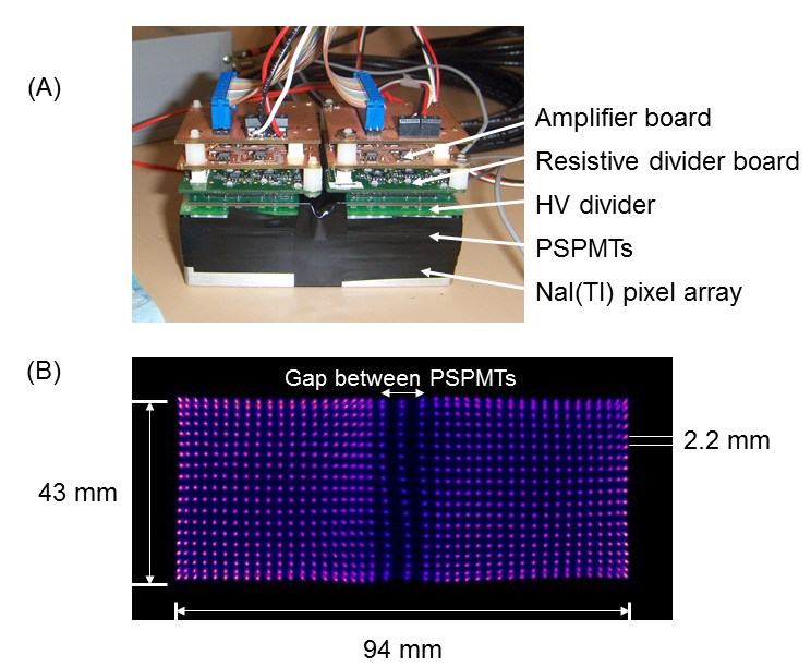
A single detector module is comprised of two position-sensitive photomultiplier tubes (PSPMTs) placed side-by-side (A). Anode row and column signals from both PSPMTs are combined through a resistive divider network to determine the X,Y position of an event. Amplification and power distribution (both high and low voltages) are also provided. Field flood illumination of the detector shows that each individual scintillation crystal in the array can be easily distinguished from its neighbor (B).
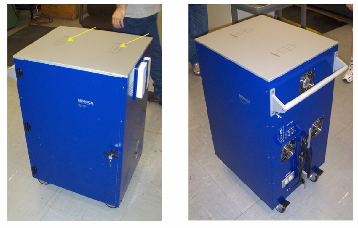
The MONICA (Mobile Nuclear Imaging Cameras) cart. The yellow arrows (left panel) point to the two upward-looking gamma cameras. A router (white box, left panel) enables wireless communication between a general-purpose laboratory Mac laptop computer and the embedded computers within MONICA. The tabletop is 62-cm wide × 55-cm deep, and 97-cm above the floor.
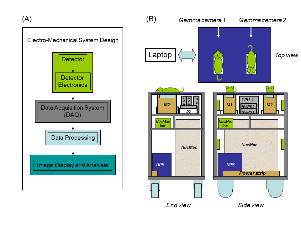
A block diagram (A) showing the basic data flow of the MONICA system. The detector and associated electronics generate analog signals from incoming gamma rays, which are then converted into digital data by the DAQ system. Data from signals outside a desired energy window are removed by a data processing algorithm. An image is formed from the remaining data, which may be further processed depending on the needs of the user. The physical layout of the MONICA system is shown on the right (B).
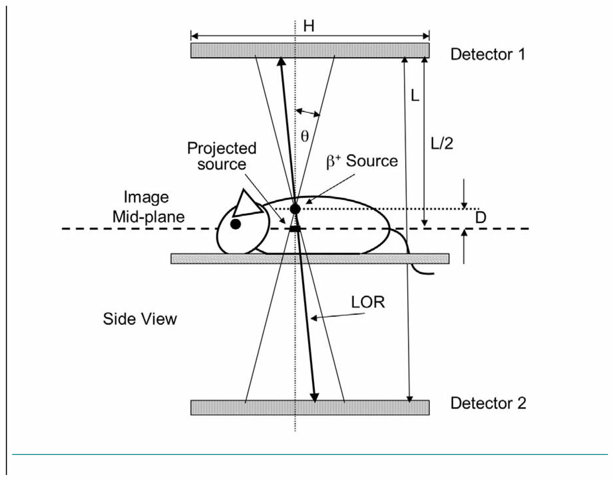
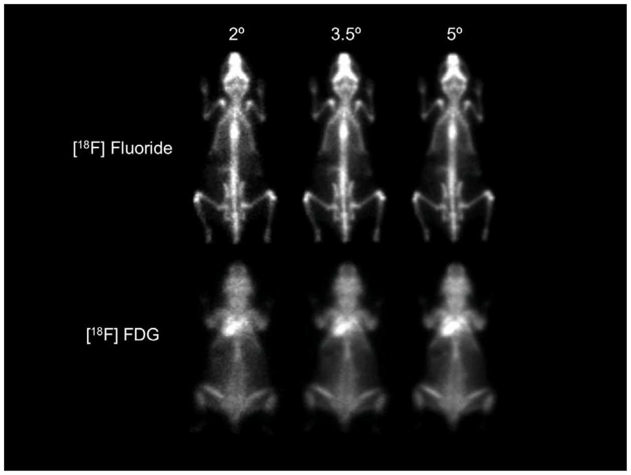
Image formation with the positron projection imager (PPI) system. The point of intersection of an activated line-of-response (LOR) with the image mid-plane is incremented by one count if the LOR lies within a small user-selected cone angle (θ) and energy window. The ensemble of all such events within cone angle θ forms a projection image of the positron annihilation distribution in the animal on the image plane. Sources above and below the image plane are ‘blurred’. The angle θ has been greatly exaggerated for clarity in this figure, and the figure objects and distances are not to scale.
Positron Projection Imager whole-body images of the distribution of [18F] fluoride (31-g animal) and [18F] FDG (25-g animal) for three different cone angles and an energy window of 250–650 keV. These images suggest that a cone angle of 3.5 degrees is a reasonable tradeoff between resolution and sensitivity.