Microfabricated Polymeric Vessel Mimetics for Cancer Cell Culture

Share:

EmailFacebookLinkedInXWhatsAppShare

Collaborators

Laboratory of Cell Biology, Center for Cancer Research, NCI
Laboratory of Cell Biology, Center for Cancer Research, NCI (formerly with IDEAS)
Biomedical Engineering & Physical Science Shared Resource Program, NIBIB
Mathematical & Statistical Computing Laboratory, Office of Intramural Research, CIT

Project Brief

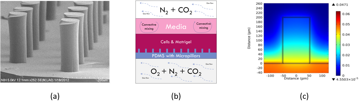
Studies evaluating potential chemotherapeutics and multi-drug resistance in cancer cells have been established using in vitro 2D culture and in vivo animal models. However, neither method accurately recapitulates human tissue or the course of tumorigenesis. 3D cell cultures better simulate the in vivo environment for tumor growth, but is currently limited by insufficient mass transport of oxygen due to a lack of vasculature.

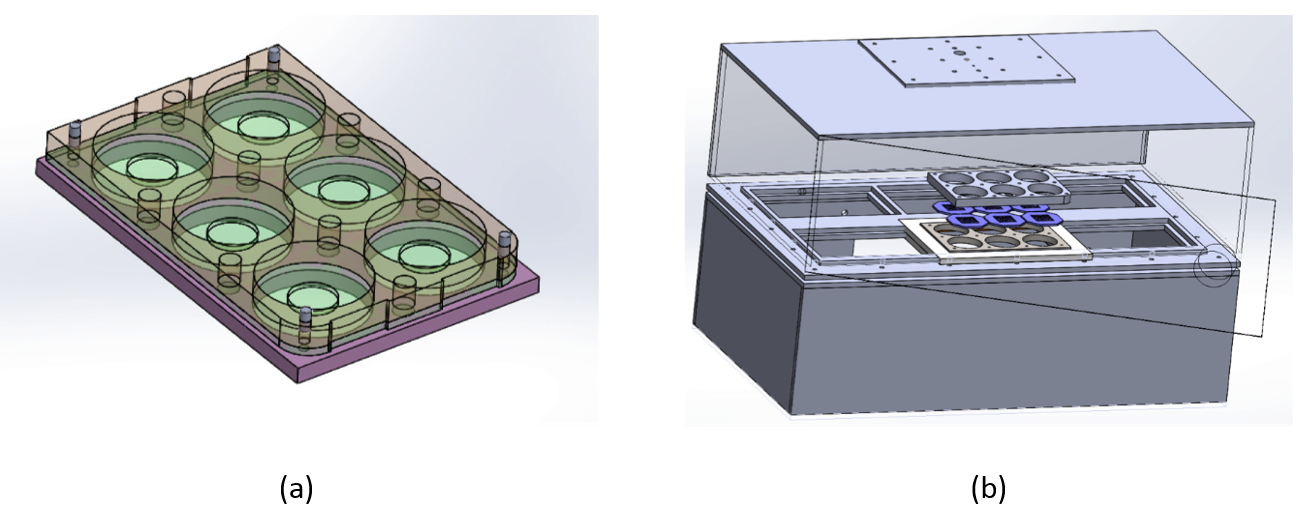
An interdisciplinary team from NCI, NIBIB, and CIT SPIS laboratories has developed microfabricated synthetic mimetics of vasculature to provide oxygen to 3D cultured tumors and other tissues. A second generation prototype, consisting of a bioreactor chamber system accommodating a six-well plate format, has been recently developed, undergone preliminary characterization, and tested under a limited number of cell culture conditions. The bioreactor setup allows for multiple 6-well plates increasing experimental throughput, and in particular:  to enable multiple parallel culture conditions under controlled hypoxia and to expand the potential application of the system to a variety of problems, including drug-screening studies. 


SEM image of micropillars, bioreactor conceptual design, O2 gradient modeling

(a) Scanning electron microscope image of micropillars cast from molds.  Pillars are ~240 microns tall with a diameter of ~82 microns.  (b) Schematic showing the conceptual diffusion of O2 through oxygen-permeable micropillars to oxygenate surrounding cells suspended in the Matrigel basement membrane extract. (c) Modeling of local O2 gradient during culture around a single micropillar, O2 concentration in mM.


3D models of bioreactor system

(a) Multi-well sandwich assembly design with an oxygen-permeable transwell membrane in the middle layer to hold the cell cultures.  Each well contains the micropillar membrane with cells, Matrigel, and media. (b) Bioreactor chamber prototype accommodating up to six multi-well plates.  A mixture of O2, N2, and CO2 flow into the lower chamber while a mixture of N2 and CO2 flow into the upper chamber.  O2 diffuses up through the micropillar membrane into the cell cultures.


Staining of tumor organoid removed from bioreactor system

Pimonidazole staining of a tumor organoid (OVCAR8-dsRed2 cells) removed from a micropillar membrane after a 7 d culture in the bioreactor system. The bottom chamber of the bioreactor was maintained at 3% O2. The location of the micropillar passing through the organoid is circled. Hypoxia level increases with distance from micropillar.


Tech Transfer

Patent application:  Microfabricated Polymeric Silicone Hydrogel Vessel Mimetics