
Collaborators
Project Brief
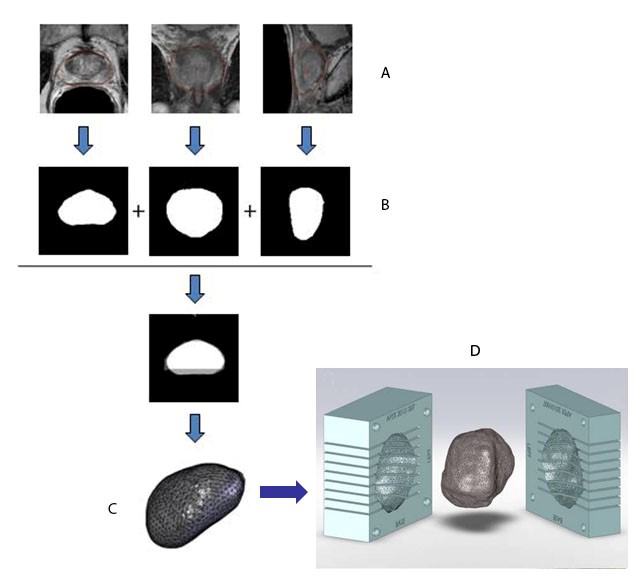
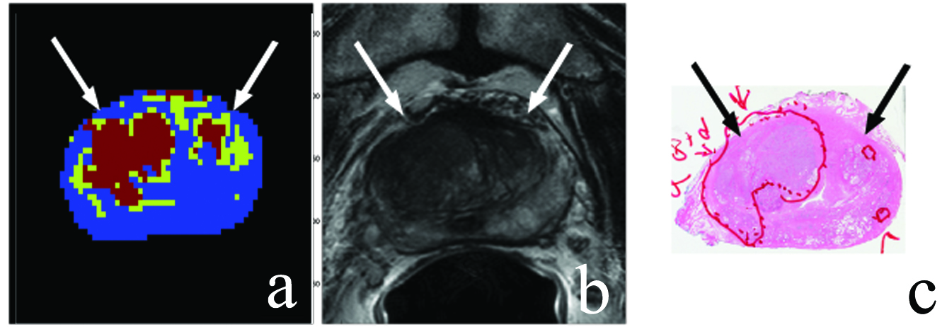
The NCI Molecular Imaging Program develops innovative methods of localizing prostate cancer based on in vivo multiparametric MRI. SPIS contributes to this effort in two areas. (1) Assisted in the design, implementation, and validation of individualized MR-based molds to facilitate the correlation of the multiparametric MRI imaging with downstream histopathology. Achieving sufficient MR/histopathology image correlation (i.e., 3D alignment) is critical when evaluating the efficacy of new prostate imaging protocols as potential cancer diagnostics. During the initial mold development phase, SPIS 3D-printed ~130 patient-specific molds. Recently, SPIS has resumed the mold printing (2-5 surgeries/week), and is now responsible for automating the process of creating the mold 3D model files from the MRI imaging files. (2) Developing an unsupervised multi-characteristic framework for prostate cancer localization using diffusion-weighted magnetic resonance imaging (DW-MRI).
More specifically, the potential single imaging modality localization method consists of combining a number of unique parameters from different models that characterize underlying tissue structure estimated from DW-MRI signal attenuation. The process of combining unique parameters from multiple models allows for the reduction of false positive and false negative voxels compared to using a single parameter, such as only apparent diffusion coefficient or kurtosis. This is due to the fact that theoretically a ‘positive-if-all-positive’ selection process favors higher specificity.
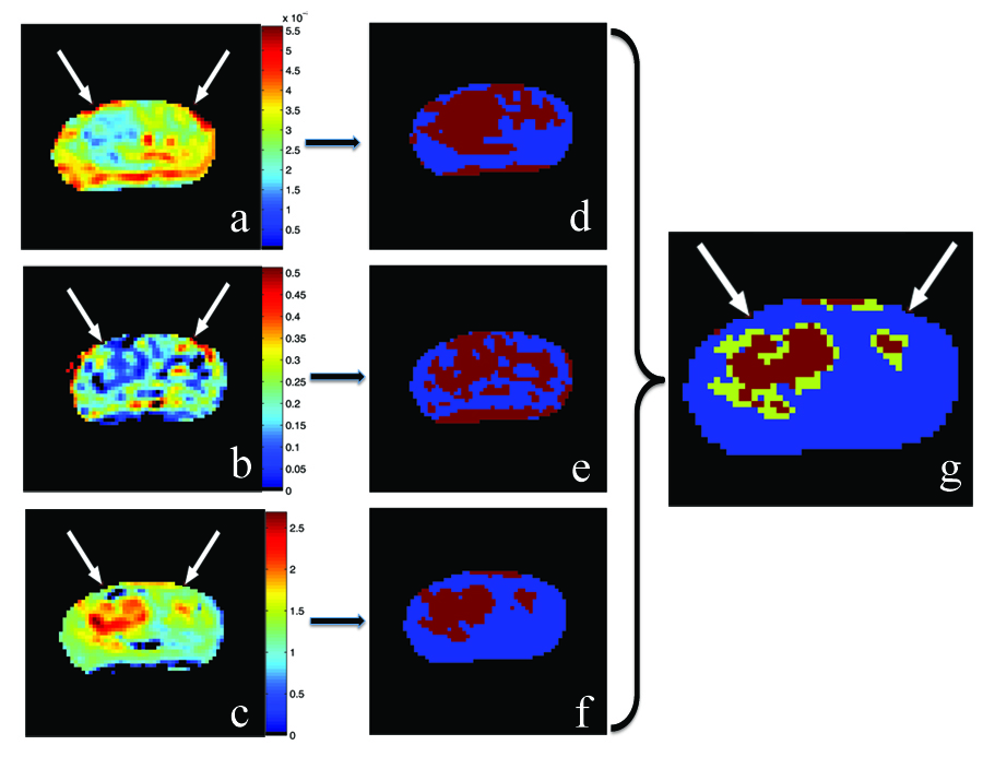
- Application of an unsupervised multi-characteristic framework for intermediate-high risk prostate cancer localization using diffusion-weighted MRI
- Atlas based AAM and SVM model for fully automatic MRI prostate segmentation
- Decision support system for localizing prostate cancer based on multiparametric magnetic resonance imaging
- 11C-Acetate PET/CT Imaging in Localized Prostate Cancer: A study with MRI and Histopathologic Correlation
- Correlation of magnetic resonance imaging tumor volume with histopathology
- A Step Towards Personalized Medicine: Use of a Patient-Specific MRI based Prostate Mold for Validation of Multi-parametric MRI in the Localization of Prostate Cancer
- Multiparametric 3T prostate magnetic resonance imaging to detect cancer: histopathological correlation using prostatectomy specimens processed in customized magnetic resonance imaging based molds
- A method for correlating in vivo prostate magnetic resonance imaging and histopathology using individualized magnetic resonance-based molds
- Novel Method for Correlating MRI and Histopathology Using Patient-Specific MR-based Molds (unavailable)
- Pathological Correlation of Radiological-Detected Prostate Cancer Lesions by a Novel Technique (unavailable)
Awards
2012 NIH Director’s Award: Design and implementation of a method for correlating in vivo prostate MRI and histopathology using individualized MR-based molds resulting in a standard-of-care at NIH