Collaborators
Project Brief
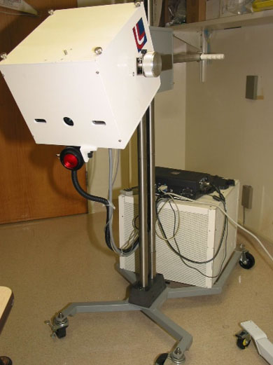
SPIS collaborated with the NICHD Program in Physical Biology to develop a clinical multispectral imaging system. The instrument acquires images of the target area in up to six different spectral bands (700 nm, 750 nm, 800 nm, 850 nm, 900 nm, and 1000 nm). Images at these wavelengths are input into both a mathematical optical skin model and a reconstruction algorithm to quantify the percent of blood in a given area, as well as the percent of blood that is oxygenated in the tissue and surrounding vasculature. The methodology has been applied to assess vascular Kaposi’s sarcoma lesions and surrounding tissue before and during experimental therapies. Results indicate that these imaging techniques are able to provide quantitative and functional information about tissue changes during experimental drug therapy, and investigate progression of disease before changes are visibly apparent; suggesting value as an alternative or complementary clinical assessment. SPIS is responsible for developing the instrumentation software for control and data acquisition.
SPIS is currently in the process of developing an upgraded multispectral imaging system. The new instrument incorporates a CCD camera with higher sensitivity and increased spectral detection range. Six fixed optical filters will be replaced with an acousto-optic filter, which can be set to any wavelength between 600 nm and 1100 nm, providing more flexibility in optical wavelength selection for specific applications.

(a)
(b)
(a) First generation portable multispectral imaging system. (b) Block diagram of multispectral imaging system. Research efforts are focused on the red and near infrared spectrum because these wavelengths penetrate the epidermis.
