
- What are medical X-rays?
- What are the benefits of medical X-rays and how are they used?
- Are there risks?
- How does NIBIB support research in X-ray technology?
What are medical X-rays?
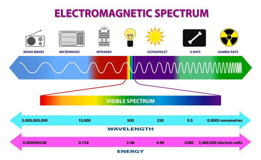
X-rays are a form of electromagnetic radiation, similar to visible light. Unlike light, x-rays have higher energy and can pass through most objects, including the body. Medical X-rays are used to generate images of tissues and structures inside the body. If X-rays traveling through the body pass all the way through to an X-ray detector on the other side of the patient, an image will be formed that represents the “shadows” formed by the objects inside of the body.
Digital radiography detectors are advanced imaging devices that capture X-ray images digitally, offering significant advantages over traditional screen-based systems, which they have largely replaced. A common type of digital X-ray is mammography that’s used for screening and diagnosis.
A digital X-ray detector can create high-resolution mammograms to detect breast cancer and other breast diseases. It works by placing a patient’s breast on a flat digital detector support plate and compressing it with a parallel plate called a paddle. An X-ray machine produces a small burst of X-rays that pass through the breast to a detector located on the opposite side. Breast density can play a role in the detection of abnormalities or cancer, which is why women with dense breasts may require additional imaging. Read more on the National Cancer Institute’s webpage on mammograms.
Computed tomography (CT) is another type of X-ray device, where a motorized X-ray source and detector quickly rotate around the patient. The detector sends signals to the machine’s computer to generate cross-sectional images or “slices.” These slices can give a clinician more detailed information than conventional X-ray scans due to the removal of overlapping tissues. Learn more about CT here.
What are the benefits of medical X-rays and how are they used?
Medical X-rays have screening, diagnostic, and therapeutic benefits. These include:
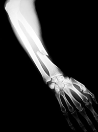
- Detecting a range of health problems including broken teeth and/or bones, bone changes or abnormalities, herniated discs in the spine, scoliosis and other spine curvature conditions, osteoarthritis, infections (e.g. pneumonia or other lung infections), certain tumors and other abnormal masses, kidney stones, calcifications, foreign objects, or dental problems.
- Screening for breast cancer: older screen film and more recent digital mammograms can detect tumors that tend to appear as regular or irregular-shaped masses and tiny bits of calcium called microcalcifications. While the latter is usually benign, specific patterns of microcalcifications could indicate the presence of cancer.
- Showing the inside of the body in real time with a series of X-rays that use a continuously monitoring digital detector. Health care professionals use the technology to diagnose conditions and to help guide medical procedures such as stent or catheter placement.
- Treating cancer: X-rays and other types of radiation given at high levels can be used to destroy cancerous tumors and cells by damaging their DNA. The radiation dose used for treating cancer is much higher than the radiation dose used for diagnostic imaging. Therapeutic radiation can come from a machine outside of the body or from a solid, liquid, or gaseous radioactive material that is placed and/or inhaled into the body or injected into the blood stream.
Are there risks?
When used appropriately, the diagnostic benefits of X-ray scans, including mammograms, significantly outweigh the risks. X-ray scans can diagnose potentially life-threatening conditions such as blocked blood vessels, bone degradation, and infections. However, X-rays produce ionizing radiation—a form of radiation that can harm living tissue. This is a risk that increases with the number of exposures that occur over the lifetime of an individual. However, the risk of developing cancer from radiation exposure is generally considered to be very small.
An X-ray in a pregnant woman poses no known risks to the baby if the area of the body being imaged isn’t near the abdomen or pelvis. Children are more sensitive to ionizing radiation and have a longer life expectancy than adults, which puts them at greater risk of developing cancer from such radiation. Parents may want to ask the X-ray technologist or doctor if their machine settings have been adjusted for children.
How does NIBIB support research in X-ray technology?
A new type of X-ray detector could improve coronary artery calcium scoring: Cardiovascular diseases are the leading cause of death globally. To assess whether there is plaque build-up (atherosclerosis) in the heart, a test is used to measure the amount of calcium in the coronary arteries. The calcium score is determined using a CT scan. However, the use of calcium scoring has been limited in general because of concerns about the low spatial resolution of CT, the radiation dose, and patient costs. To address these limitations, researchers are developing a new type of X-ray detector (a dual-layer flat panel) that could improve calcium scoring.
Developing a low-cost dual energy X-ray source for computed tomography: A dual-energy X-ray source for CT scans is increasingly used for various clinical tasks and has the potential to provide significantly more information than a regular CT. However, the higher cost of this technology has limited its widespread adoption. NIBIB-funded researchers are developing a new dual-energy X-ray source that could potentially be less expensive and perform better. The technology could be applied to imaging of a patient’s arms and legs, the face and head, as well as image-guided radiation therapy and interventional radiology.
Updated September 2025