
Project Brief
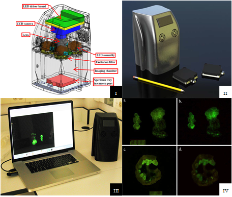
In collaboration with the NCI Molecular Imaging Program (MIP), SPIS designed and implemented a novel portable fluorescence imaging system which has proven to be a valuable asset in the clinical translation of molecular imaging probes. Molecular imaging probes allow surgeons and endoscopists to visualize tumor tissue which normally would not be visible to the naked eye. While the initial testing and validation of the molecular probes can be done in MIP laboratories with a variety of commercial imaging systems, the clinical validation and translation may require a collaboration with outside hospitals in order to gain access to patients with the specific tumor type being targeted. Offsite collaborations pose several problems, including regulations regarding transportation of the human tissue, regulations regarding patient privacy, and requirements that the probe be applied to the tissue shortly after resection. In order to overcome these restrictions, SPIS designed a small, portable fluorescence camera which NCI researchers could easily transport to the offsite location; eliminating the need for tissue transport, simplifying patient privacy concerns by reducing the number of people who need access to the tissue, and allowing the probes to be applied in an optimal timeframe. As there were ongoing patient surgeries as part of an existing collaboration, MIP researchers required a solution as quickly as possible. In approximately one month, SPIS staff leveraged our in-house design expertise to quickly develop the full system, including: custom LED lighting for excitation, optical system for probe excitation and emission, camera integration, support electronics, and 3D-printed enclosure. The completed camera system was validated using control and animal samples. While the initial collaboration studied ovarian cancer, the imaging system has been a critical component in other cancer studies by our NCI collaborator from the University of Tokyo
Tech Transfer
Duplicated system for use by Dr. Urano, an NCI collaborator from the University of Tokyo