
Collaborators
Project Brief
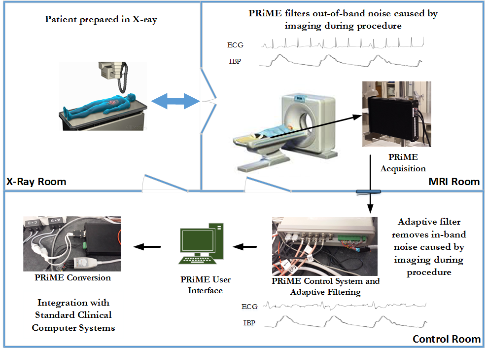
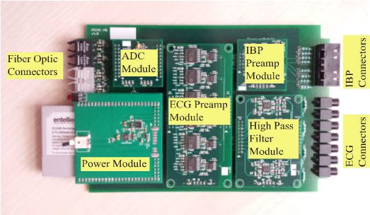
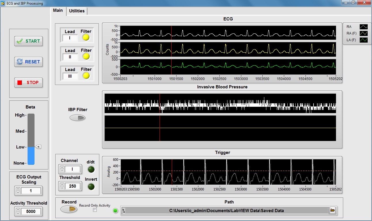
A multidisciplinary intramural team led by CIT in collaboration with the NHLBI Cardiovascular Intervention Program developed and implemented a fundamental electronics and signal processing tool facilitating MRI catheterization procedure innovation. As an alternative to surgical techniques in the treatment of cardiovascular disease, NHLBI is developing innovative techniques in cardiovascular catheterization with the use of real-time MRI. The use of MRI greatly reduces the patient’s exposure to ionizing radiation from X-ray imaging, which is typically used in catheterization procedures. During these procedures, the cardiology staff must continuously monitor the patient’s physiological signals (i.e., ECG and IBP (invasive blood pressure)) for both patient safety and diagnostic purposes. Due to the lack of commercial solutions, SPIS staff developed a high fidelity physiological signal recording system that operates in the hostile (e.g., electromagnetic interference) environment of MRI, which allows physicians to safely navigate catheters through the heart using MRI. The Physiological Recording in MRI Environment (PRiME) system includes custom electronics which are resilient to MRI-induced noise (e.g., filtering, digitization, data transmission, fabrication methods tailored to mitigate the effects of a noise-rich environment), an adaptive filter software algorithm which removes additional noise, and an interface to a commercial hemodynamic recording system which allows the cardiology staff to use their existing workflow. In a license-free form, SPIS is publicly disseminating the PRiME system design documents for other research hospitals to duplicate the PRiME system, allowing more research groups to contribute to the field of MRI-guided cardiovascular interventions. With NHLBI, SPIS deployed the PRiME system to the Children’s National Medical Center in Washington, D.C., where it has been successfully used in over 40 pediatric procedures. Within NHLBI at the NIH Clinical Center, the PRiME system has been used in over a half dozen adult procedures and numerous animal experiments.
Tech Transfer
- Public Distribution of Design Files on GitHub
- System duplication and deployment to Children’s National Medical Center, Washington D.C.
- System duplication and deployment to Children’s Health, Dallas, TX
Awards
- 2016 NIH Director’s Award: The Development of Physiological Recording in MRI Environment (PRiME) for the Implementation of Radiation-Free MRI-Guided Right Heart Catheterization
- 2015 NHLBI Director's Award: Outstanding Clinical Research Award For the Implementation of Radiation -Free, MRI-Guided Right Heart Catheterization in Pediatric Patients