
Collaborators
Project Brief
Energy-driven proton pumps are of major importance for energy-transduction in living systems. The respiratory chain of animals, as well as single cellular organisms, uses energy released from electron transport to oxygen to form an electrochemical gradient for protons, which is then used to synthesize ATP, a critical element in intracellular energy transfer. To understand the specific, atomic-level, details of the electrogenic transport of protons across the membrane, our NHLBI collaborator uses spectrophotometry to study the rapid kinetics of the simple, photon-driven, proton pump bacteriorhodopsin (BR). Information obtained from this proton pump should help in understanding more complex systems such as mammalian cytochrome oxidase, an important enzyme in the respiratory process.
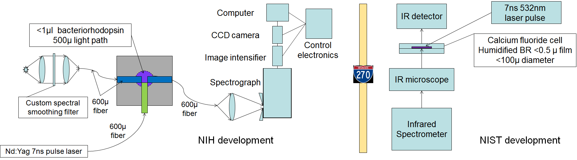
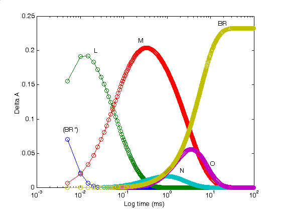
Continuing a collaboration which has spanned decades, SPIS engineers developed a high-speed scanning spectrometer system to obtain time-resolved absorption spectra on the kinetic reaction mechanisms. The spectrometer system used a highly parallel approach to simultaneously acquire 96-channels covering the visible spectrum from 350 nm to 900 nm. Using advanced singular value decomposition methods, our collaborators have conducted studies which support the parallel cycle model of the BR photocycle instead of a single photocycle model which is favored by many researchers.
A collaboration with NIST led to further development to coordinate experiments from a commercial IR spectrometer (Bruker), allowing data from both systems to be combined and analyzed as a complete data set covering both IR and visible wavelengths. The combined infra-red and visible spectrum optical spectroscopy produced kinetic structural information that characterizes each step of the photocycle. The success of the initial studies lead to further work to produce single crystals of the BR membrane protein. The overall goal of this project is to characterize the BR crystal using similar established approaches in concert with time resolved X-ray diffraction to obtain structural information at the atomic level, thus enabling an understanding of protein conformational changes that result in electrogenic transport of protons across the membrane.
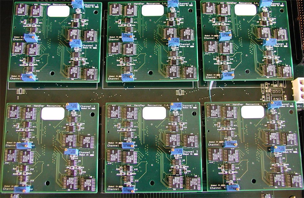
A multi-IC collaboration (NHLBI, NIBIB, NIST, CIT) developed a next generation system based on a commercial image intensifier, spectrograph, and CCD camera. The new system was designed to measure the extremely low light levels of the single membrane BR crystals as well as provide seamless coverage of the full spectrum. No instrument capable of performing such studies has been previously described. The new system will be used for: 1) quality control to verify whether a membrane protein functions the same in the crystal as in situ in the membrane, 2) as a guide to alter the crystallization procedure to produce crystals more closely related to in situ function, and 3) to produce valid crystals to be used in time-resolved X-ray crystallography (in order to obtain isolated atomic structures for each intermediate in the proton-pumping procedure so that the overall process can be better understood).
Tech Transfer
Interagency Collaboration. Deployed system to National Institute of Standards and Technology laboratory in support of long-standing collaboration with NHLBI investigator.