

Guofeng Zhang
, Ph.D.
Biologist
The Electron Microscopy Unit of the BEPS provides state-of-the-art instrumentation, training, and services. The unit collaborates on projects using immuno-electron microscopy and electron tomography. Training on specimen preparation, including cryo-techniques, can also be provided. Please find a full list of services and pricing in the lab resources section below.
Transmission Electron Microscopy (TEM)
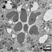
- Fully automated FEI Tecnai T12 transmission electron microscope
- High-sensitivity 2k x 2k CCD camera for digital imaging and transfer of data across network directly to users’ laboratories.
- Automated electron tomography, and energy-dispersive x-ray spectroscopy
- Immuno electron microscopy
Preparative Techniques for TEM
- Standard fixation, embedding and sectioning (Leica EM UC6 microtome)
- High-pressure fixation (BalTec HPM 010)
- Freeze-substitution (Leica EM AFS 1/2)
- Pre- and post-embedding immunolabeling, cryosectioning (Tokyuasu technique) (Leica Ultracut UCT/FCS cryomicrotome)
- Negative staining of macromolecular assemblies
Scanning Electron Microscopy
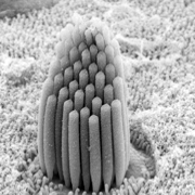
- Fully automated Hitachi S-4800 field-emission scanning electron microscope (FESEM) provides tool for determining 3D surface morphology of cells and biomaterials
- Users can be trained to collect SEM images independently
- Sputter coater (Balzers) for specimen preparation
- Critical point dryer (EM Sciences)
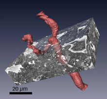
Serial Block Face Scanning Electron Microscopy (SBF-SEM)
- Fully automated Zeiss Sigma/Gatan 3View Serial block face SEM provides 3D imaging of tissues (up to 300 microns across) at the nanoscale
- Visualization software for 3D rendering of biological structures
- Specialized specimen preparation techniques for SBF-SEM of tissues and isolated cells
Location of Electron Microscopy Unit
Building 13 on the NIH campus
Pricing
- Tecnai T12 TEM (BEPS staff operation) $150 per hour
- Tecnai T12 TEM (client operation) $80 per hour
- Zeiss SIGMA/Gatan 3View SBF-SEM $100 per hour
- Hitachi S-4800 (client operation) $100 per hour
- EM Sample Embedding $300 for 1-5 samples
- EM Sample Sectioning $80 per sample
- EM Immunostaining $400 per set
32451441[uid] OR 32726643[uid] OR 22517616[uid] OR 34857844[uid] OR 29543442[uid] OR 34401678[uid] OR 26020550[uid] OR 32375181[uid] OR 31061173[uid] OR 30401752[uid] OR 28557263[uid] OR 24466366[uid] OR 30420913[uid] OR 22425023[uid] OR 23228423[uid] OR 27520728[uid] OR 34531522[uid] OR 28470979[uid] OR 21210706[uid] OR 25684354[uid] OR 30418749[uid] OR 21055473[uid] OR 26301492[uid] OR 25501370[uid] OR 26565809[uid] OR 26474409[uid] OR 22540867[uid] OR 22064945[uid] OR 25648146[uid] OR 26928972[uid] OR 21366330[uid] OR 19718033[uid]