
Science Highlights
January 4, 2016
Dr. Sanjay Jain is an avid Star Trek fan who began his recent NIH Wednesday Afternoon Lecture with a nod to the cult TV and film franchise. The quintessential medical device in the starship Enterprise sick bay is the “tricorder”—a hand-held scanner waved over a patient to detect injury and heal all manner of maladies.
“Unfortunately, we don’t have tricorders yet, but we have imaging machines and the concept is pretty straightforward,” he said.
“You inject a tracer that goes to the bug or whatever metabolic process you want to measure, it’s labeled with something that emits energy, and you have machines that will then tell you where the energy is coming from, which will correlate with the disease process.”
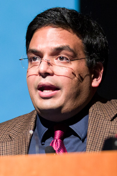
Jain is associate professor of pediatrics and international health at Johns Hopkins University. He also directs JHU’s Center for Infection and Inflammation Imaging Research. He receives support for his work from the National Institute of Biomedical Imaging and Bioengineering, the NIH Common Fund, NHLBI and NIAID. He titled his talk “Bugs, Drugs and Star Trek.”
The technologies that might be needed for tricorder-like medical care may not be light years away, according to Jain. In fact, the Food and Drug Administration has approved three molecular tracers that, with positron emission tomography (PET) imaging, will detect the presence of amyloid plaques in the brains of living patients, a biomarker for Alzheimer’s disease. But he said that there is not a similar tracer yet for infectious diseases. So his team, which investigates how modern imaging methodology can be applied to research on infectious disease and to patient care, has taken up this research challenge.
Jain’s team is conducting experimental work in animals using a combination of computed tomography (CT) and PET scanning techniques to track the behavior of infectious disease in real time. The group’s investigations include imaging studies in tuberculosis, which typically, but not exclusively, attacks the lungs. The laboratory also is developing imaging techniques to detect other bugs, such as the family of bacteria that includes E. coli.
“It is not easy to do imaging for TB because it is a biosafety-level 3 pathogen, which means that it spreads by the aerosol route,” Jain said. To prevent transmission of TB, he devised an air-tight, unbreakable and transparent container to bring infected mice to an imaging facility, accumulating important data to advance understanding of ways to monitor the disease in animals and develop novel bacteria-specific imaging tracers. There are almost 10 million cases of TB worldwide each year, many of which are resistant to drug therapies.
The week prior to his talk, Jain’s group published a case in Lancet Infectious Diseases of a 2-year-old girl who contracted a form of tuberculosis known as XDR TB, or extensively drug-resistant TB. He presented the case as a vignette of the life-and-death circumstances that are a kind of reality check for the work of his laboratory.
The child—now 5 years old and in remission—was seen in the hospital after returning from a 3-month visit to India. She experienced persistent fever, but the doctors’ initial tests could not pinpoint the cause. A chest X-ray revealed a suspicious lung spot, so the doctors tested lung secretions and forged ahead with preemptive treatment for TB. It took 12 weeks for the tests to conclusively identify her case as drug-resistant TB.
With no fast, reliable way to monitor how bacteria responded to the new treatment, the team used CT to rapidly track the response to therapy. The scans were a low-radiation, child-friendly adaptation of that imaging technique, which could become the norm for pediatric CT imaging.
Current imaging methods, such as CT, magnetic resonance imaging and ultrasound, cannot reliably distinguish infection from cancer or other processes. With imaging probes that can specifically label infections, imaging can become clinically useful, Jain said. He presented a variety of promising research studies in which imaging scans with several tracers could detect infections in mice. His lab is hoping to develop a pipeline of imaging probes to identify, locate and monitor a wide range of pathogenic bacteria within laboratory animals and eventually translate those techniques to patients.
Jain emphasized the importance of developing new tools to improve patient care. Contrasting his work with the challenge faced by cancer researchers, who attempt to use imaging to distinguish between healthy and cancerous cells, he says that detecting microbes is relatively easy.
“Microbes—bacteria, prokaryotes and fungi—have had billions of years of evolution and have very unique metabolic pathways and structures,” he said, noting that cell walls in these organisms are completely unique. “There is nothing like that present in the human body,” he said. “So I think it’s a low-hanging fruit.”