Artificially causing – or inducing – labor is becoming increasingly common, yet this practice comes with risks and its level of success is difficult to foresee. But now, new research may offer a way to help predict outcomes and improve the process.
Researchers at the University of Arkansas for Medical Sciences (UAMS) have devised a non-invasive method of accurately measuring the electrical activity of uterine muscles. The results of a recent clinical study, published in the journal Current Research in Physiology, show that signals measured in pregnant patients prior to induction are strongly tied to whether their labor lasted less or more than 24 hours. The authors indicate that physicians could use the method to learn how patients might respond to induction and use the information to develop more effective strategies for labor and delivery.

If a pregnant person or their baby’s health would benefit from beginning labor sooner, health care providers may recommend inducing the process through medication or other means. Without any reliable monitoring instruments, physicians heavily rely on physical examinations and their experience to inform induction strategies.
Taking this approach, time spent in labor varies greatly from patient to patient and there is also a risk of excessive doses of induction medication causing harm.
“Sometimes women will sit there for 36 hours after being induced and nothing’s happening,” said Hari Eswaran, Ph.D., professor of obstetrics and gynecology at UAMS. “We wanted to know when the uterus is prepared for labor. If we capture a physiological signature beforehand that indicates the chances of successful induction, that information can be used to personalize the approach.”
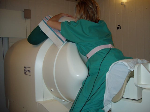
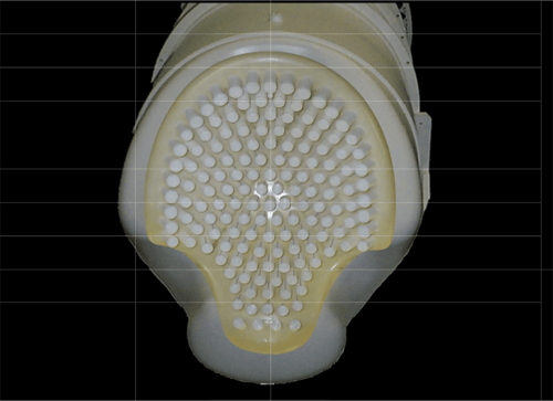
In search of telling cues, Eswaran and his co-authors previously developed the Superconducting Quantum Interference Device (SQUID) Array for Reproductive Assessment, or SARA for short.
Gauging the electrical activity of various parts of the body through techniques such as electromyography, or EMG, is routine in the clinic. However, electrical signals from the uterus weaken by the time they reach the surface of the skin, making them particularly challenging to detect.
Instead of directly measuring electrical activity, SARA takes a back door.

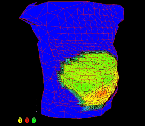
Electrical current, whether passing through power cables or the muscles of the human body, always generates a magnetic field around it. And if you can detect the field, then you can work backwards to calculate the electrical current that produced it, Eswaran explained. This is the approach the researchers are taking with SARA’s array of 151 biomagnetic sensors.
In the new study, the authors analyzed measurements taken from 27 patients that were between 37 and 42 weeks pregnant. Each rested their abdomen on SARA’s sensors, which recorded the magnetic activity produced by the uterus prior to induction. The authors converted the magnetic signals into metrics related to the electrical activity, such as power. After noting how long it took for each patient to deliver, the authors examined their data and picked up on a significant pattern.
Patients that delivered their babies within 24 hours after induction exhibited four times as much uterine electrical power across entire recordings than those in labor for longer.
While none of the patients were experiencing contractions while SARA recorded signals, the results suggest that those with higher uterine electrical power were more prepared for labor, Eswaran explained.

Equipped with a tool that could help predict responsiveness to induction, physicians would be better informed in devising strategies tailored to individual patients. With further research, SARA could become such a tool, potentially providing guidance on when to administer induction medication and in what doses, which would lead to safer and more efficient labor and delivery.
“NIBIB provided grant funding over 15 years ago to support the development of the novel computational methods used in this clinical trial to predict labor contractions with SARA,” said Grace C.Y. Peng, Ph.D., director of the NIBIB program in Mathematical Modeling, Simulation and Analysis. “It is so rewarding to see this project demonstrate the translational potential of computer models to improve the quality of care in women’s health.”
This research was funded in part by a grant from NIBIB (R01EB007264).
This Science Highlight describes a basic research finding. Basic research increases our understanding of human behavior and biology, which is foundational to advancing new and better ways to prevent, diagnose, and treat disease. Science is an unpredictable and incremental process—each research advance builds on past discoveries, often in unexpected ways. Most clinical advances would not be possible without the knowledge of fundamental basic research.
Study reference: Sarah Mehl et al. Assessing uterine electrophysiology prior to elective term induction of labor. Current Research in Physiology (2023). DOI: 10.1016/j.crphys.2023.100103
