
Precise mapping of functional networks aims to improve brain surgeries and research
Improvements in brain sensing technologies have allowed clinicians to perform increasingly complex surgeries and enabled researchers to map the signals of the brain that control feeling, movement, and thought. Now a multidisciplinary research team has designed, fabricated, and tested the latest in brain-probing technologies that promises to unlock the brain’s functional networks in unprecedented detail.
To obtain maps of functional signals, brain probing technologies often consist of grids of electrodes placed directly on the brain like a small net. This type of grid with sensors to record signals is known as electrocorticography (ECoG). During surgery to remove tumors or epileptic regions, signals emanating from the grid guide the removal of diseased tissue while helping surgeons avoid damage to surrounding healthy tissue. In research, grids placed on the brain in animals and humans are used to record and map the signals that carry out specific functions such as reaching and grasping.
Despite these remarkable achievements, unraveling the complexity of the brain has only begun.
To address the challenge of mapping the brain with ever greater precision a team of engineers, medical researchers and surgeons have harnessed the power of nanotechnology to build grids that can record signals from the human brain with significantly greater accuracy than current technologies. The project is led by electrical engineering professor Shadi Dayeh at the University of California San Diego Jacobs School of Engineering and includes team members from UC San Diego, Massachusetts General Hospital, and Oregon Health & Science University.
The new nanotech-enabled grids are comprised of several thousand sensors—compared to the current ECoG grids which typically have between 16 and 64 sensors.
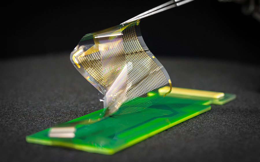
The technology is called a platinum nanorod grid (PtNRGrid). The PtNRGrid creates 100-fold more precise brain maps because the tiny nano sensors can be densely packed—placed just one millimeter apart—compared to current grids with sensors spaced at one-centimeter intervals.
“A central problem in electronics and sensors is that when placed too close together one sensor interferes with the signal of the other,” explained Moira Bittman, Ph.D., director of the program in Biorobotic Systems at the National Institute of Biomedical Imaging and Bioengineering (NIBIB), which co-funded the research. “The team has used elegant engineering and fabrication techniques that allow these powerful sensors to function precisely while in extremely close contact. It is a great example of the type of innovations we support at NIBIB that are poised to deliver significant breakthroughs in research and medicine.”
The team tested the new technology in rats and in humans undergoing brain surgeries.
A PtNRGrid about the size of a kernel of corn containing 1024 sensors was placed on a small region of a rat brain. Tiny puffs of air were used to stimulate sensory areas on the rat such as whiskers and skin. When a puff of air contacted a single whisker, the grid picked up the electrical signal in the area of the brain that controls the sensations of that exact whisker. The PtNRGrid produced a map of the brain identifying the area of the brain connected to each of the 16 individual whiskers spaced just fractions of a millimeter apart—a level of detail not possible with previous technologies.
A PtNRGrid the size of a postage stamp containing 1024 or 2048 nanosensors was also used in experiments in humans. The patients had given permission for the research studies while undergoing surgery for tumors or to remove brain regions causing epileptic seizures.
The 1024 sensor PtNRGrid identified the brain region controlling sensation in each of the five fingertips in the test subjects as well as the regions controlling a grasping motion. Remarkably, the grid was able to pick up the distinct brain signals involved in the planning stage of the motion, the actual motion, and after the completion of motion—signals that occur just millionths of a second apart.
In tests in a patient with epilepsy using 2048 sensors, the PtNRgrid was able to identify the location of brain areas discharging epileptic waves with millimeter accuracy as well as test waves the researchers induced in different areas of the brain. The millimeter resolution of the areas identifying where the signals originated were superior to the centimeter resolution that can be obtained with currently used technologies. This level of accuracy could allow a surgeon to remove diseased tissue just millimeters away from healthy tissue rather than using a margin of centimeters, thus sparing more function.
{"preview_thumbnail":"/sites/default/files/styles/video_embed_wysiwyg_preview/public/video_thumbnails/-7ggs6e2UXI.jpg?itok=BBjQgNIq","video_url":"https://youtu.be/-7ggs6e2UXI","settings":{"responsive":1,"width":"854","height":"480","autoplay":0},"settings_summary":["Embedded Video (Responsive)."]}
“We are encouraged and extremely excited about the possibilities offered by this new technology,” explained Dayeh. “We estimate that hundreds of thousands of individuals would benefit from the significantly more accurate brain surgeries that will be possible using the PtNRGrids. In addition, our process for manufacturing the grids is similar to that used to make the screens on televisions, phones and computers, which allows us to efficiently add functional utility to the grids.”
By “functional utility” Dayeh explains that the team is working to produce cost-effective grids that will provide even more precise and useful information to surgeons while performing these extraordinarily difficult procedures.
The study was published in Science Translational Medicine1. The work was supported by the NIH award no. NIBIB DP2-EB029757 to S.A.D., BRAIN® Initiative awards R01NS123655-01 to S.A.D. and UG3NS123723-01 to S.A.D., and NIDA R01-DA050159 to S.A.D.; NSF award no. 1728497 to S.A.D. and CAREER no. 1351980 to S.A.D.; and an NSF Graduate Research Fellowship Program no. DGE-1650112 to A.M.B.