
Single-cell variations can indicate if treatment is working, or time to change course
Understanding the variability in individual cells of a tumor can be a valuable guide to the choice and course of treatment. NIBIB-funded researchers used photoacoustic imaging for rapid measurement of oxygen consumption rate of individual cells from breast tumors—information that can help guide treatment strategies, monitor results, and allow for adjustments over the course of treatment.
Treating solid tumors is challenging because as they grow, each cell in the tumor takes on different genetic and metabolic characteristics, which can make them more or less susceptible to particular treatments. This makes killing every cancer cell in a tumor extremely difficult.
“Getting as much information as possible about a tumor to allow for effective treatment is an ongoing goal of cancer researchers,” explains Behrouz Shabestari, Ph.D., director of the Program in Optical Imaging at the National Institute of Biomedical Imaging and Bioengineering, which funded the study. “The photoacoustic technique developed by these researchers was previously used to non-invasively look into human tissues for abnormalities such as tumors. Now they have advanced the technology to detect differences in each cell in the tumor, which moves us closer to developing personalized therapies based on the specific metabolic characteristics of each person’s tumor cells.”
Dr. Shabestari is speaking of the senior investigator of the study, Lihong Wang, Ph.D., Bren Professor of Medical and Electrical Engineering, and his colleagues at the California Institute of Technology. The technique is photoacoustic imaging, in which harmless pulses of laser light hit the target, creating vibrations that return as a detailed image of the target in the form of sound, similar to ultrasound images.
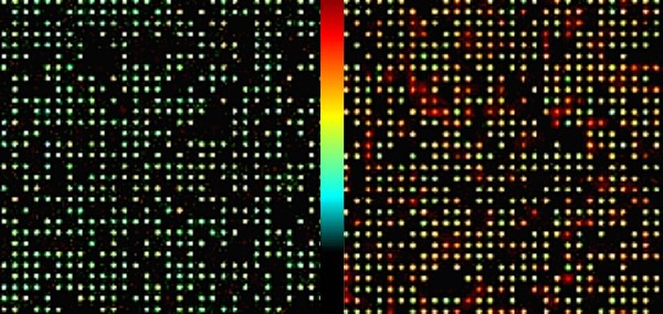
In this case, Dr. Wang and his group have modified their technique to determine how much oxygen is being used by each cell of a tumor that has been individually isolated in a tiny well on a laboratory plate. Each cancer cell is sitting in its own well surrounded by blood filled with oxygen-carrying hemoglobin. Each cell is using up different amounts of the oxygen in the well at a different rate, which reveals differences in the metabolism of each cell.
To measure oxygen levels with lasers and sound, the researchers modified their technique to develop single cell metabolic photoacoustic microscopy (SCM-PAM). When the laser hits each well, it causes the hemoglobin in the well to vibrate. The technique works because hemoglobin that is bound to oxygen vibrates differently from hemoglobin without oxygen.
The varying vibrations that come from each well ultimately create different colors that provide a measurement of how much oxygen has been used up by each individual cancer cell. The less oxygen left in the well of a cell, the higher the metabolic rate of that cell.
Another important aspect of this new technology is the speed at which it can measure the metabolic rate of a batch of cells isolated from a tumor. Using SCM-PAM, Wang and his team were able to measure the rate of oxygen consumption of 3,000 cancer cells in just 15 minutes, which is about one hundred times faster than a method that uses individual oxygen sensors in each well that is extremely difficult and expensive to build and operate.
The technique was tested on a cancer cell line and on breast cancer cells obtained by biopsies from patients. In both cases the cancer cells had higher oxygen consumption than normal cells, which correlates with the faster growth of the cancer cells. The test also identified individual rates of oxygen consumption for each cell.
“This new technique allows us to evaluate an important aspect of cancer cells, which is how much the metabolic rate is increased compared to normal cells from the same patient,” explained Wang. “We also observed a wide range of different metabolic activity in the population of cancer cells from the same tumor.”
The researchers expect this new tool to be extremely valuable for cancer research as well as in the clinical setting. “One good example of the value of the technique is that we can look at the metabolic rate of the cancer cells from a patient before and after treatment,” says Wang. “If the rate is reduced it is a good indicator that the therapy that was chosen is working. If not, it gives and opportunity to switch to a different therapy much more rapidly than is possible by current means, which can be as long as several months to determine whether a tumor is shrinking.”
The study was reported in the April edition of Nature Biomedical Engineering.1 The work was supported by an NIH Director’s Pioneer Award DP1 EB016986 through the National Institute of Biomedical Imaging and Bioengineering, an NIH Director’s Transformative Research Award R01 CA186567 through the National Cancer Institute, and a grant from the National Science Foundation.