
Study in rabbits uses biodegradable piezoelectric film to aid in growth of new cartilage
Osteoarthritis—a painful condition that results from the deterioration of the cartilage in our joints—affects millions of people worldwide. This persistent problem has been challenging to address, as joint repair techniques, such as cartilage grafts, have extensive limitations, and current tissue regeneration treatments have limited use in human patients. To combat this issue, NIBIB-funded researchers are developing an implantable, biodegradable film that helps to regenerate the native cartilage at the site of damage. Their study, performed in rabbits, could be an initial, important step in the establishment of a new treatment for this common condition.
“While most tissue engineering methods rely on growing artificial tissues in the lab, this regenerative medicine technique recruits the body’s own cells to facilitate healing of the damaged cartilage,” said David Rampulla, Ph.D., director of the division of Discovery Science & Technology at NIBIB. “This preclinical study, while early, describes a promising approach to regenerate damaged cartilage, which has the potential to benefit those with osteoarthritis and other forms of cartilage damage.”
Traditional surgical treatments for cartilage defects can involve a cartilage graft to replace the damaged tissue. These cartilage grafts are either taken from somewhere else in the body (from a non-weight bearing joint, for example) or are taken from a donor. But both of these options can have potential complications: Grafts taken from another joint can cause additional damage and scarring, while grafts taken from a donor are in limited supply and may be rejected by the body. Other treatments under investigation include growing cartilage in a tissue dish, but so far, this process is time-consuming and has a variety of limitations.
"Rather than treating osteoarthritis by using a cartilage graft at the site of injury, we wanted to develop a therapy that stimulates the native, damaged cartilage to regenerate itself,” explained Thanh Nguyen, Ph.D., an associate professor at the University of Connecticut. And it so happens that cartilage can regenerate when it is exposed to a small electrical charge.
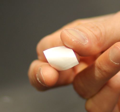
But instead of hooking up wires or a battery to a damaged joint, Nguyen and colleagues are using a different approach—inside the damaged cartilage, they are implanting biodegradable films with piezoelectric properties that generate an electrical charge when compressed. That way, when the joint bears weight from movement or exercise, the film produces an electrical charge that recruits and stimulates cells to regenerate cartilage at the area of damage. Results from this approach were recently published in Science Translational Medicine.
What is piezoelectricity? Piezoelectricity is the ability of a specific material to generate an electrical charge when subjected to mechanical forces, such as pressure. Examples of objects that use piezoelectric components include gas lighters, musical greeting cards, and ultrasound transducers.
In their study, the researchers first evaluated how well their piezoelectric films could enhance chondrogenesis, or cartilage formation, of stem cells grown in the lab. The cells were grown on the surface of the piezoelectric film, which was inside of a chamber that could be subjected to controllable pressure. The researchers programed the chamber to apply a specific amount of pressure for 20 minutes per day for 14 days. They found that these conditions resulted in increased production of components that make up cartilage, such as collagen and glycosaminoglycans (a type of polysaccharide).
Nguyen explained how the electrical charge stimulated new cartilage growth: “We found that cells grown on the piezoelectric scaffolds under pressure had enhanced secretion of an important growth factor involved in wound healing, known as TGF-beta,” he said. What’s more, they found that the charged surface of the piezoelectric film attracted significantly more fibronectin, a protein involved in tissue healing, compared with a non-piezoelectric film. “We believe that the electrical charge generated by our films can both recruit proteins important for tissue regeneration and can stimulate nearby cells to secrete factors involved in wound healing,” Nguyen said.
The researchers next evaluated their piezoelectric films in rabbits with damaged knee joints, which is a way to model osteoarthritis. The animals were divided into several categories: rabbits with implanted piezoelectric films, rabbits with implanted films that were not piezoelectric, and rabbits without any implanted films. Within each category, the animals were further categorized into groups that either participated in an exercise regimen—20 minutes of hopping on a treadmill per day—or did not participate in such exercise, after one month of recovery following surgery.
Then Nguyen and colleagues analyzed how well the damaged cartilage had healed. They found that the rabbits with implanted piezoelectric films that participated in the exercise regimen had the most cartilage regeneration; in fact, the entire defect in these damaged joints were refilled with new tissues. Analysis by computed tomography (CT) revealed that rabbits in this group also had enhanced bone regeneration around the knee joint.
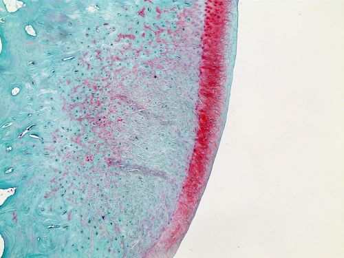
When the researchers evaluated the cartilage tissues under a microscope, they confirmed that animals treated with implanted piezoelectric films and an exercise regimen had the most improved regeneration, compared with the other groups. These animals had abundant chondrocytes (cartilage cells) and collagen protein in the joint that was treated. “Treatment with the piezoelectric film coupled with exercise resulted in a beautiful layer of cartilage that looks like the native tissue in our models,” said Nguyen.
While the results are promising, Nguyen stressed that this work is still not ready to be evaluated in humans. “The materials that we are using in our films are very safe, and the manufacturing process is very scalable, so in that sense, our technology is very translational,” he said. “However, important next steps will include the evaluation of our films in larger animals, with larger joint loads, before studies can begin in human patients.”
This study was funded by a grant from NIBIB (R21EB024787) and partially by a grant from NIAMS (for National Institute of Arthritis and Musculoskeletal and Skin Diseases; R21AR078744).
Study reference: Yang Liu, Godwin Dzidotor, Thinh T. Le, Tra Vinikoor, Kristin Morgan, Eli J. Curry, Ritopa Das, Aneesah McClinton, Ellen Eisenberg, Lorraine N. Apuzzo, Khanh T. M. Tran, Pooja Prasad, Tyler J. Flanagan, Seok-Woo Lee, Ho-Man Kan, Meysam T. Chorsi, Kevin W. H. Lo, Cato T. Laurencin, and Thanh D. Nguyen. Exercise-induced piezoelectric stimulation for cartilage regeneration in rabbits. Science Translational Medicine, 2022; 14 (627). DOI: 10.1126/scitranslmed.abi7282