Image Library
Axial Slices of the Brain
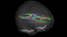
The primary eigenvector of the diffusion tensor in these two axial slices indicates the fiber orientation at each voxel. A transparent brain surface rendering provides a sense of position and scale.
Ronin Protien in an Oocyte
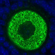
Image of an oocyte (unfertilized egg cell) with an abundance of a protein called “Ronin” which appears to play a significant role in the pluripotency of embryonic stem cells.
Source: Karen Hirschi, Baylor College of Medicine
Magnetic Resonance Elastography
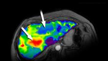
An image produced by a new, noninvasive way to diagnose liver fibrosis using a new technique called MR Elastography which uses sound waves to determine tissue firmness. Doctors are able to see inside the liver without biopsies. The red indicates diseased tissue while the blue indicates healthy tissue.
Source: Richard Ehman, Mayo Clinic
Instant Mobile Cervical Cancer Diagnosis
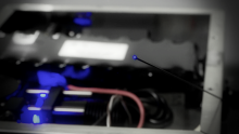
Image of a portable, fiber optic microscope that non-invasively characterizes and diagnoses precancerous and cancerous cells. Both low cost and battery-powered, this point-of-care device is ideally suited to low resource settings and facilitates immediate outpatient therapy.
Source: Rebecca Richards-Kortum, Rice University
Engineering Human Livers
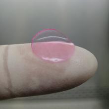
Image of a mini bioengineered human liver that can be implanted into mice. This capability enables researchers to investigate how human livers metabolize drugs, to test susceptibility to toxicity, and to demonstrate species-specific responses that typically do not show up until clinical trials.
Source: Sangeeta Bhatia, MIT
Bioengineered Tissue Scaffold
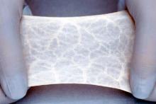
A biomaterial made from pigs' intestines which can be used to heal wounds in humans. When moistened, the material, which is called SIS, is flexible and easy to handle.
Source: Stephen Badylak, University of Pittsburgh.
Targeting Cells for Drug Delivery
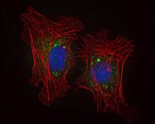
Image of nanoparticles entering the cell and releasing their drug payload. The cell skeleton is shown in red, the cell nucleus in blue. The green color inside the cell demonstrates that the nanoparticles entered the cell.
Source: Omid Farokhzad, Brigham and Women’s Hospital
Self-administered Vaccine Patch
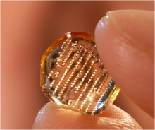
Image of a new vaccine patch that can be self-administered and does not produce biohazardous waste. The microneedles in the patch inject the vaccine into the epidermis layer and then dissolve.
Source: Mark Prausnitz, Georgia Tech
Building an Artificial Ovary
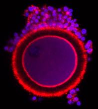
Image of a baboon oocyte (unfertilized egg cell) surrounded by granulosa cells which help in the development of the oocyte.
Source: Northwestern University
